
Spinose Ear Tick
Ixodes kingi
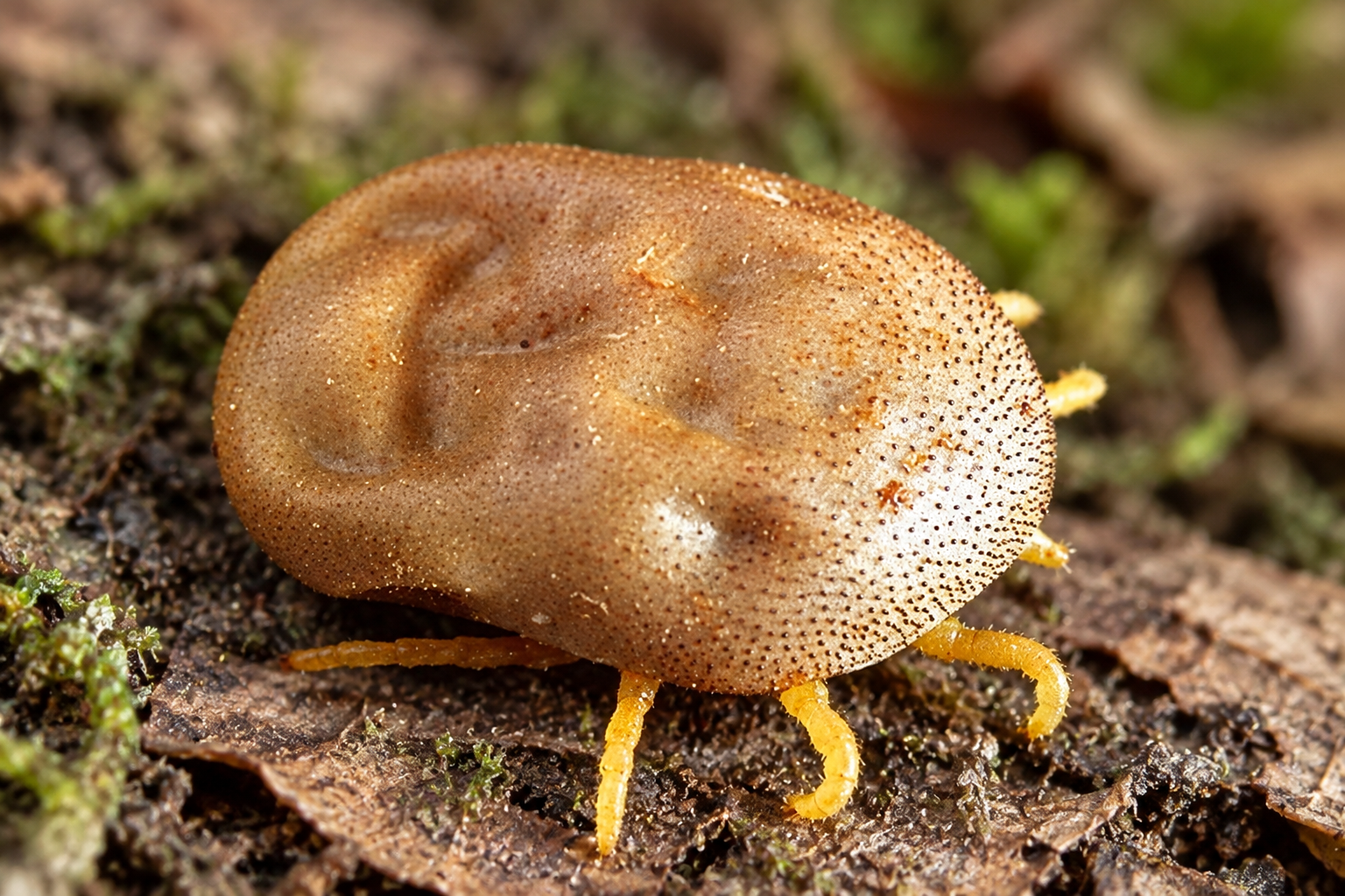
Primarily found on deer (especially mule deer and black-tailed deer), but can also be found on other mammals such as elk, livestock, dogs, and Occasionally humans.
Most commonly found in forested and wooded areas, brushy habitats, and regions where deer are common, particularly in the western United States and parts of western Canada.
Can carry and potentially transmit pathogens such as Anaplasma phagocytophilum (the bacterium that causes anaplasmosis) and Babesia odocoilei (a Babesia species).
 Identification & Key Features
 Identification & Key Features
 The Spinose Ear Tick looks very different from most hard ticks.
As a soft tick, it:
- Lacks a hard dorsal scutum
- Has a soft-bodied appearance rather than the rigid shield typical of hard ticks
- Changes considerably in appearance between life stages
- Has parasitic larvae and nymphs
- Has nonfeeding adults
The distinctive spiny appearance is most obvious in the later nymphal stage, rather than in the adult.
Adult Female
Adult females:
- Have 8 legs
- Lack a hard scutum
- Have a granular, soft-tick body surface
- Are generally grayish, blue-gray, or brownish
- Have reduced, nonfunctional feeding mouthparts
- Do not feed
- Live entirely off-host
- Mate in the environment
- Lay eggs in protected environmental locations
Adults of the species typically measure approximately 4–8 mm long, with females generally larger than males.
Adult females survive using nutrients retained from the final nymphal blood meal.
Published estimates of egg production vary substantially. Texas A&M describes females laying up to approximately 500 eggs over 2–4 weeks, while Merck Veterinary Manual reports that females may deposit as many as approximately 1,500 eggs under some conditions. Reproductive output therefore should not be represented by a single universal egg number.
Adult Male
Adult males:
- Have 8 legs
- Lack a scutum
- Have a granular, soft-bodied appearance
- Have reduced, nonfunctional feeding mouthparts
- Do not feed
- Live entirely off the host
- Mate with females in protected environmental locations
Unlike adults of many hard-tick species, adult male and female Spinose Ear Ticks are not normally found attached to animals.
An actively feeding O. megnini found inside an ear is therefore an immature tick—a larva or nymph—not an adult.
Nymph
Nymphs are the most distinctive and commonly recognized stage.
They:
- Have 8 legs
- Are generally gray to light brown or blue-gray
- Lack a scutum
- Have well-developed feeding structures
- Develop conspicuous spine-like setae
- Often have a characteristic rounded, constricted, or peanut-shaped appearance
- Live and feed within the external ear canal
- Develop through two nymphal instars
The pronounced spines of the later nymphal stage give the species its common name:
Spinose Ear Tick.
The parasitic nymphal period can be prolonged. Oklahoma State University reports that nymphs may feed repeatedly over approximately 30–200 days, while other veterinary sources describe the combined larval and nymphal parasitic period as lasting several weeks to several months.
Larva
Larvae:
- Have 6 legs
- Are extremely small
- Are generally pale, gray, or light brown
- Have functional feeding mouthparts
- Are the principal host-seeking stage
- Climb vegetation, feed bunks, or other elevated objects
- Wait for contact with a passing animal
- Crawl toward the head after reaching the host
- Enter the external ear canal
- Attach and feed
- Eventually molt into the first nymphal stage while remaining on the same host
Texas A&M describes newly hatched larvae ascending vegetation to await a host before moving into the inner portion of the outer ear.
Other Identification Features
Important identification characteristics include:
- Soft tick — family Argasidae
- No scutum
- Larvae have 6 legs
- Nymphs and adults have 8 legs
- Later nymphs have conspicuous spine-like setae
- Nymphs often have a peanut-shaped appearance
- Larvae and nymphs feed primarily within the ear canal
- Adults have reduced, nonfunctional feeding mouthparts
- Adults do not feed
Using a magnifier can help make the tick’s spines, legs, body texture, mouthparts, and other identification features easier to see.
Distinguishing It From Other Ticks
The Spinose Ear Tick is especially distinctive when its life stage, body structure, and feeding location are considered.
Spinose Ear Tick — Otobius megnini
- Soft tick
- No scutum
- Later nymphs have conspicuous spines
- Immature stages specialize in feeding inside the external ear canal
- Adults do not feed
- Feeding stages normally remain on a single host
Typical Hard Ticks — Family Ixodidae
- Have a scutum
- Adults normally feed
- Mouthparts generally project forward and are visible from above
- Most species are not specialized for prolonged development deep within the ear canal
Because ticks inside an ear may be partly hidden by wax, debris, swelling, or inflammation, careful examination may be necessary.
Geographic Distribution & Habitat
Geographic Distribution & Habitat The Spinose Ear Tick is native to the Americas and is especially associated with dry, warm, or relatively low-rainfall environments.
In North America, it is particularly associated with:
- The western United States
- The southwestern United States
- Parts of the south-central United States
- Mexico
- Western Canada, particularly British Columbia
It is also well established in areas such as Oklahoma, where it can be an important livestock parasite.
Its documented North American range extends from western Canada through the United States and Mexico. Although especially associated with arid and semiarid western regions, records occur beyond its historically recognized southwestern range.
The species has also been transported with livestock to other parts of the world, including portions of:
- Central America
- South America
- Southern and southeastern Africa
- India
- Madagascar
- Hawaii
- The Galápagos Islands
- Other regions where introductions have occurred
Common Habitats
Habitat depends strongly on life stage.
On the Host
Larvae and nymphs occur primarily:
- Deep within the external ear canal
- In protected portions of the outer ear
- Around the inner portions of the pinna and ear opening
The ear canal provides a protected environment where immature ticks can remain and feed for an extended period.
Off the Host
Adults and eggs occur in protected environmental locations such as:
- Barns
- Stables
- Livestock pens
- Around feed bunks
- Beneath boards
- Cracks and crevices
- Beneath debris
- Around fences, logs, and sheltered livestock areas
Adults live entirely off-host because they do not feed.
Life Cycle & Seasonal Activity
Life Cycle & Seasonal Activity The Spinose Ear Tick has an unusual life cycle:
Egg → 6-Legged Larva → First Nymphal Instar → Second Nymphal Instar → Nonfeeding Adult
The species uses a single host for its parasitic feeding stages.
A larva attaches to an animal and normally remains associated with that same host while developing through both nymphal instars. Only after the final nymphal feeding period does the tick leave the host.
Eggs
Adult females lay eggs off the host in protected locations.
Eggs may be deposited:
- Beneath boards
- Around feed bunks
- In cracks and crevices
- In barns or livestock shelters
- In other protected environmental sites
Texas A&M reports that eggs generally require approximately three weeks to hatch under suitable conditions, although incubation time varies with temperature and other environmental conditions.
Larvae
After hatching, larvae:
- Have 6 legs
- Crawl upward onto vegetation or structures
- Wait for a suitable host
- Transfer onto a passing animal
- Move toward the head
- Enter the external ear canal
- Attach and feed
The larva then molts into the first nymphal instar without leaving the host.
First Nymphal Instar
The first nymph:
- Has 8 legs
- Remains within the ear
- Reattaches and continues feeding
- Continues developing on the same host
- Molts into the second nymphal instar
Second Nymphal Instar
The second nymph:
- Has 8 legs
- Develops the characteristic spinose appearance
- Continues feeding within the ear canal
- May remain parasitic for weeks to months
- Eventually completes its final blood meal
After completing the parasitic stage:
Second-stage nymph → detaches from host → drops into environment
The tick then molts into a nonfeeding adult.
Adults
Adults:
- Live entirely off-host
- Have reduced, nonfunctional feeding mouthparts
- Do not feed
- Mate in protected environmental locations
- Depend on nutrient reserves obtained during the final nymphal feeding stage
Merck Veterinary Manual reports that adults may survive for almost two years despite never taking an adult blood meal.
After mating, females lay eggs and the cycle begins again.
Seasonal Activity
Spinose Ear Tick infestations can occur throughout the year.
However, immature-stage parasitism is often most prominent during:
Late winter and spring
Texas A&M reports that although immature ticks may cause problems year-round, parasitism of hosts is typically greatest during late winter and spring. Merck similarly describes larval and nymphal feeding as occurring predominantly during winter and spring.
Oklahoma State University reports that larval activity in Oklahoma begins increasing around March, illustrating that exact seasonal timing varies geographically.
For a national website, the clearest summary is:
Spinose Ear Ticks can infest animals throughout the year, but feeding-stage infestations are often greatest during late winter and spring. Exact timing varies with geography, climate, host availability, and local environmental conditions.
Diseases & Health Risks
Diseases & Health Risks The primary health problems associated with the Spinose Ear Tick result from:
- Direct feeding
- Pain
- Ear-canal irritation
- Inflammation
- Tissue injury
- Secondary infection
Its major importance is therefore direct parasitism and veterinary injury rather than established transmission of infectious disease.
Ear Canal Irritation & Inflammation
Spinose Ear Tick larvae and nymphs feed within the external ear canal.
Infested animals may develop:
- Ear pain
- Severe irritation
- Inflammation
- Restlessness
- Frequent head shaking
- Head rubbing
- Drooping ears
- Sensitivity around the ears
- Excessive wax or debris
- Discomfort when moving the head
Current Merck Veterinary Manual guidance identifies O. megnini infestation as a cause of external ear-canal irritation in horses, cattle, sheep, goats, deer, rabbits, dogs, and cats.
Secondary Ear Infections
Heavy or prolonged infestation can damage the ear canal.
Feeding wounds combined with:
- Inflammation
- Ear wax
- Tick feces
- Shed tick skins
- Moisture
- Tissue debris
can create conditions favorable for secondary microbial infection.
Texas A&M specifically notes that tick-infested animals can develop secondary infection because of feeding wounds and the accumulation of feces and cast exuviae.
Secondary infection may contribute to:
- Worsening inflammation
- Ear discharge
- Tissue damage
- Sloughing of damaged tissue
- Additional pain
Oklahoma State University specifically reports secondary bacterial infection and sloughing of ear tissue in affected animals.
Hearing Impairment
Large numbers of ticks can cause severe inflammation and may obstruct or substantially affect the ear canal.
Heavy infestations have been associated with:
- Severe irritation
- Inflammation
- Ear-canal blockage
- Hearing impairment or deafness
Infested cattle may also develop a characteristic “flop-eared” appearance associated with discomfort and damage from infestation.
Tick Paralysis & Neurologic Effects — Animals
Tick paralysis has been reported in association with Otobius megnini infestation in animal hosts, although it is not the species’ primary or most common clinical effect.
Merck Veterinary Manual includes O. megnini among tick species associated with paralysis in animals. Tick paralysis results from biologically active substances associated with tick feeding rather than from transmission of an infectious pathogen.
Horses
In horses, Spinose Ear Tick infestation has also been associated with painful intermittent muscle cramping.
Affected horses can develop:
- Severe episodic muscle cramps
- Abnormal muscle contractions
- Episodes resembling colic
- Falling when stimulated in severe cases
Treatment of the underlying ear-tick infestation has been associated with resolution of the syndrome.
Secondary Maggot / Screwworm Infestation
Severely damaged or inflamed ear tissue can attract flies.
Flies may lay eggs in affected tissue, resulting in myiasis or maggot infestation.
Secondary infestation by screwworm larvae has been reported in animals with severe Spinose Ear Tick infestations.
Human Health Risk
Humans can be parasitized by Otobius megnini.
Human cases can involve larvae or nymphs feeding inside the external ear canal, producing a condition known as otoacariasis.
Possible effects include:
- Ear pain
- Irritation
- Inflammation
- A sensation of movement or blockage
- Local tissue injury
- Ear-canal swelling
- Possible secondary complications
Human infestation is uncommon compared with infestation of livestock and wildlife.
Because very small larvae may be difficult to visualize and ticks may attach deep within the ear canal, otoscopic, microscopic, or endoscopic examination can sometimes be necessary for complete evaluation.
Important Disease-Transmission Clarification
The Spinose Ear Tick is not currently established as a vector of a major human tick-borne disease in the United States.
This distinction is important.
Merck Veterinary Manual notes that O. megnini has been found carrying or infected with agents associated with:
- Coxiellosis / Q fever
- Tularemia
- Colorado tick fever
- Rocky Mountain spotted fever
However, Merck specifically states that the tick’s ability to transmit these pathogens to hosts is unknown. Texas A&M likewise lists no known disease pathogen associated with established transmission by this species.
Therefore:
Finding a pathogen in a tick is not the same as demonstrating that the tick is a competent natural vector capable of transmitting that pathogen to a host.
For a public-facing U.S. page, the accurate conclusion is:
The Spinose Ear Tick is primarily important because of direct ear-canal parasitism and veterinary injury. Transmission of a major human tick-borne pathogen by this species has not been established.
Important Lyme Disease Clarification
The Spinose Ear Tick is not a recognized Lyme disease vector.
In the United States, Lyme disease bacteria are transmitted to people primarily by:
- Ixodes scapularis — Blacklegged Tick
- Ixodes pacificus — Western Blacklegged Tick
CDC specifically identifies these Ixodes ticks as the U.S. vectors responsible for human Lyme disease transmission.
Finding Otobius megnini should therefore not be interpreted as evidence that this particular tick transmitted Lyme disease.
Important to Know
The greatest health significance of Otobius megnini is direct parasitism rather than demonstrated pathogen transmission.
Important consequences can include:
- Painful feeding
- Ear-canal irritation
- Inflammation
- Secondary infection
- Hearing impairment in severe infestations
- Loss of condition in heavily infested livestock
- Reported tick paralysis or other neurologic effects in animals
- Secondary myiasis or screwworm infestation
This makes the Spinose Ear Tick primarily a veterinary and animal-health parasite.
Bite Prevention & Protection
Bite Prevention & Protection Because this species primarily parasitizes livestock and other mammals, prevention should focus heavily on animals and their environments.
Around Livestock
Regularly inspect the ears of:
- Cattle
- Horses
- Sheep
- Goats
- Other livestock in areas where the tick occurs
Watch for:
- Frequent head shaking
- Head rubbing
- Drooping ears
- Restlessness
- Sensitivity around the ears
- Ear discharge
- Unusual odor
- Signs of pain
- Changes in hearing or responsiveness
Ticks may be located deep within the external ear canal, where they can be difficult to see without appropriate examination.
Around Barns & Stables
Because adults and eggs occur off-host:
- Keep barns and livestock areas clean.
- Reduce unnecessary debris and sheltered hiding areas.
- Address cracks and crevices where adult ticks may shelter.
- Inspect areas around feed bunks, boards, fences, and livestock resting areas.
- Follow veterinary recommendations for tick control.
- Use only appropriately labeled animal or premises treatments.
- Treat the environment when necessary as part of an integrated tick-control program.
Veterinary sources note that environmental control may be important because adult ticks live and reproduce away from the host.
For substantial infestations, consult a veterinarian or qualified livestock pest-management professional.
Human Exposure
People handling heavily infested animals should:
- Wear appropriate protective clothing when practical.
- Inspect themselves after working closely with infested livestock.
- Avoid placing fingers or unprotected instruments deep within an animal’s ear.
- Seek veterinary assistance for difficult or heavy animal ear infestations.
If You Find an Attached Tick
If a Spinose Ear Tick—or another tick—is attached to accessible human skin, remove it promptly.
CDC’s current June 9, 2026 guidance recommends removing attached ticks as soon as possible rather than delaying removal while waiting to see a healthcare provider.
How to Remove a Tick
- Use clean fine-tipped tweezers.
- Grasp the tick as close to the skin’s surface as possible.
- Pull the tick away from the skin with steady, even pressure.
- Do not twist or jerk the tick.
- If mouthparts remain in the skin and can be removed easily with tweezers, remove them. If they cannot be removed easily, leave them alone and allow the skin to heal.
- Dispose of the tick safely.
- Thoroughly clean the bite area and your hands.
- Check the rest of your body for additional ticks.
Do not attempt to make an attached tick detach by using:
- Petroleum jelly
- Nail polish
- Heat
- Other substances or chemicals
These approaches should not replace prompt mechanical removal.
Important Ear-Canal Precaution
A tick located deep inside a person’s ear canal should not be treated like an easily accessible tick attached to ordinary skin.
Attempting blind or forceful removal with tweezers, cotton swabs, or other objects can:
- Injure the ear canal
- Push the tick or debris deeper
- Damage the eardrum
- Make complete removal more difficult
Clinical reports of human otoacariasis commonly describe removal under direct visualization with appropriate instruments, followed by examination of the ear canal and eardrum.
Seek medical care for a tick located deep within the ear canal, particularly if it cannot be clearly and safely reached.
Deep ear-canal infestations in pets or livestock should likewise be evaluated by a veterinarian.
Tick Testing
CDC generally does not recommend commercial testing of removed ticks to guide medical treatment decisions.
A positive result does not prove that transmission occurred, while a negative result can provide false reassurance. Treatment decisions should not be based on commercial tick-testing results.
For O. megnini specifically, pathogen testing is even more difficult to interpret because transmission of a major human tick-borne pathogen by this species has not been established.
After Removal
Seek medical evaluation if you develop:
- Persistent or worsening ear pain
- Increasing redness or swelling
- Drainage
- Hearing changes
- Fever
- Rash
- Persistent irritation
- Other concerning symptoms
CDC advises seeking medical care if fever or rash develops within the weeks following a tick bite.
Tell the healthcare provider:
- That a tick was present
- When it was discovered
- Where the likely exposure occurred
Whether the exposure involved livestock or other animals
Identification Gallery
Identification Gallery - 8 legs
- Gray, brown, or blue-gray body
- Distinctive spiny surface
- Rounded or peanut-shaped body
Engorged Nymph
- Enlarged body after feeding
- Spiny surface
- Comparison with an unfed nymph
First-Instar Nymph
- 8 legs
- Smaller than the second-stage nymph
- Less noticeable spines
Larva
- Very small body
- 6 legs
- Comparison with a nymph
Engorged Larva
- Enlarged body after feeding
- 6 legs
- Comparison with an unfed larva
Adult
- 8 legs
- Soft-tick appearance
- Granular body surface
- Reduced mouthparts
Adult Spinose Ear Ticks do not feed.
Ear-Canal Infestation
Only larvae and nymphs feed. Heavy infestations may cause irritation, inflammation, infection, and possible hearing problems.
Life Cycle
Egg → 6-Legged Larva → First Nymph → Second Nymph → Leaves Host → Nonfeeding Adult → Mating & Egg Laying
Larvae and nymphs develop on the host. Adults live off-host and do not feed.
Soft Tick Comparison
Spinose Ear Tick (Otobius megnini)
- Immature stages mainly feed inside the ear
- Nymphs have a distinctive spiny appearance
- Adults do not feed
- Not considered a major human disease vector
Relapsing-Fever Soft Ticks (Ornithodoros spp.)
- Often found in nests, burrows, caves, or cabins
- Nymphs and adults may feed multiple times
- Some species transmit soft tick relapsing fever

